


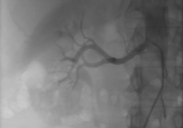
Coronary angiography is an invasive examination used to study the coronary arteries, which supply blood to the heart. Except in emergency situations, it is generally performed after non-invasive tests such as an electrocardiogram, stress test, or cardiac imaging.
This examination is the gold standard method for confirming or excluding coronary artery disease. It is also indicated before certain cardiac interventions or surgeries.
Coronary angiography involves inserting a thin catheter into an artery, most often at the wrist or sometimes at the groin, in order to inject a contrast agent visible on X-rays and visualize the coronary arteries.
The examination is performed under local anesthesia. A preliminary blood test and the placement of an intravenous line are part of the standard preparation.
Lorem ipsum dolor sit amet, consectetur adipiscing elit, sed do eiusmod tempor incididunt ut labore.
Lorem ipsum dolor sit amet, consectetur adipiscing elit, sed do eiusmod tempor incididunt ut labore.
Lorem ipsum dolor sit amet, consectetur adipiscing elit, sed do eiusmod tempor incididunt ut labore.
Lorem ipsum dolor sit amet, consectetur adipiscing elit, sed do eiusmod tempor incididunt ut labore.
Hemodynamic assessment and coronary physiology allow analysis of the actual impact of a narrowing of the heart’s arteries on blood circulation. They are indicated when the severity of a lesion is intermediate or when symptoms are not explained by the observed abnormalities.
These assessments are performed during coronary angiography using specific measurements of pressure and flow in the coronary arteries. They provide a precise functional evaluation of the lesions and their impact.
These examinations help determine whether interventional treatment is necessary, avoid unnecessary procedures, and guide management, including in cases of angina without coronary obstruction.


Lorem ipsum dolor sit amet, consectetur adipiscing elit, sed do eiusmod tempor incididunt ut labore.
Lorem ipsum dolor sit amet, consectetur adipiscing elit, sed do eiusmod tempor incididunt ut labore.
Lorem ipsum dolor sit amet, consectetur adipiscing elit, sed do eiusmod tempor incididunt ut labore.
Lorem ipsum dolor sit amet, consectetur adipiscing elit, sed do eiusmod tempor incididunt ut labore.

Intracoronary imaging is a technique performed during coronary angiography that allows examination of the inside of the heart’s arteries with great precision. It complements angiography by providing detailed information about the vascular wall and plaques.
This examination allows characterization of lesions, specification of their extent, and guidance of treatment choice and optimization. It is also used to verify proper expansion and apposition of stents, particularly in complex situations.
The use of this imaging improves the precision of interventions and contributes to optimizing short- and long-term outcomes.
Lorem ipsum dolor sit amet, consectetur adipiscing elit, sed do eiusmod tempor incididunt ut labore.
Lorem ipsum dolor sit amet, consectetur adipiscing elit, sed do eiusmod tempor incididunt ut labore.
Lorem ipsum dolor sit amet, consectetur adipiscing elit, sed do eiusmod tempor incididunt ut labore.
Lorem ipsum dolor sit amet, consectetur adipiscing elit, sed do eiusmod tempor incididunt ut labore.
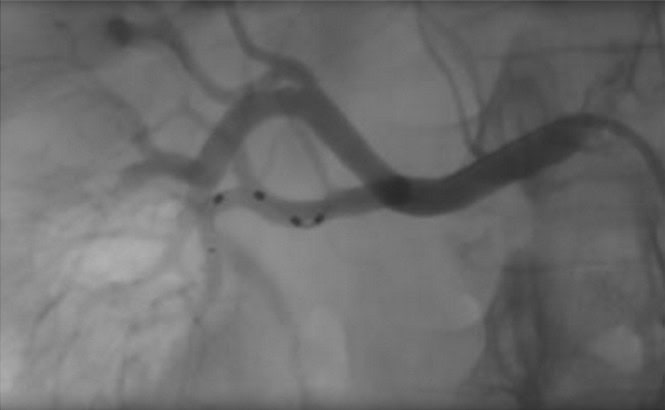
Coronary angioplasty is an intervention aimed at treating a narrowing of a heart artery. It is most often performed following coronary angiography, sometimes during the same examination.
The intervention consists of dilating the narrowed area using a balloon. Depending on the characteristics of the lesion, treatment may be completed by the placement of a stent, intended to keep the artery open, or by the use of a drug-coated balloon. The strategy is defined on an individualized basis in order to achieve the best balance between efficacy and safety.
Angioplasty is performed under local anesthesia, most often via arterial access at the wrist. Monitoring is necessary after the intervention.
Drug therapy is adapted according to the clinical situation, most often with the addition of an additional antiplatelet treatment, in order to prevent complications and optimize the long-term outcome.

Lorem ipsum dolor sit amet, consectetur adipiscing elit, sed do eiusmod tempor incididunt ut labore.
Lorem ipsum dolor sit amet, consectetur adipiscing elit, sed do eiusmod tempor incididunt ut labore.
Lorem ipsum dolor sit amet, consectetur adipiscing elit, sed do eiusmod tempor incididunt ut labore.
Lorem ipsum dolor sit amet, consectetur adipiscing elit, sed do eiusmod tempor incididunt ut labore.

Treatment of chronic total coronary occlusions (CTO) concerns heart arteries that have been completely blocked for several months. This situation can cause chest pain, shortness of breath, or exercise limitation, despite appropriate medical treatment.
The intervention is performed percutaneously, using specialized techniques to cross and then reopen the occluded artery. It relies on the use of dedicated equipment and a carefully planned strategy, adapted to each patient, in order to restore satisfactory blood circulation.
Although the principle is similar to that of conventional angioplasty, this procedure is more complex and may require advanced techniques and specific approaches. It is proposed in selected situations, when the expected benefit is clearly established.
Monitoring is necessary after the intervention to ensure proper clinical evolution. Appropriate drug therapy is implemented to prevent complications and optimize the long-term outcome.
Lorem ipsum dolor sit amet, consectetur adipiscing elit, sed do eiusmod tempor incididunt ut labore.
Lorem ipsum dolor sit amet, consectetur adipiscing elit, sed do eiusmod tempor incididunt ut labore.
Lorem ipsum dolor sit amet, consectetur adipiscing elit, sed do eiusmod tempor incididunt ut labore.
Lorem ipsum dolor sit amet, consectetur adipiscing elit, sed do eiusmod tempor incididunt ut labore.
Invasive assessment of pulmonary hypertension is performed when it is necessary to confirm the diagnosis or specify its mechanism. It allows differentiation between the various forms of pulmonary hypertension and identification of its origin.
The examination consists of right heart catheterization, performed under local anesthesia. A thin catheter is introduced through a vein in order to directly measure pressures in the right heart and pulmonary arteries, as well as vascular resistances.
This assessment is essential for establishing a precise diagnosis and guiding the choice of the most appropriate treatment. Monitoring is necessary after the procedure.

Lorem ipsum dolor sit amet, consectetur adipiscing elit, sed do eiusmod tempor incididunt ut labore.
Lorem ipsum dolor sit amet, consectetur adipiscing elit, sed do eiusmod tempor incididunt ut labore.
Lorem ipsum dolor sit amet, consectetur adipiscing elit, sed do eiusmod tempor incididunt ut labore.
Lorem ipsum dolor sit amet, consectetur adipiscing elit, sed do eiusmod tempor incididunt ut labore.

Invasive assessment of valvular heart disease is proposed when non-invasive examinations do not allow precise determination of the severity of a valvular lesion or guidance of management.
It is based on hemodynamic measurements performed during cardiac catheterization, allowing precise analysis of pressures and the function of the affected valves.
These data complement imaging examinations and contribute to defining the most appropriate therapeutic strategy, whether it involves monitoring, interventional treatment, or surgical management.
Lorem ipsum dolor sit amet, consectetur adipiscing elit, sed do eiusmod tempor incididunt ut labore.
Lorem ipsum dolor sit amet, consectetur adipiscing elit, sed do eiusmod tempor incididunt ut labore.
Lorem ipsum dolor sit amet, consectetur adipiscing elit, sed do eiusmod tempor incididunt ut labore.
Lorem ipsum dolor sit amet, consectetur adipiscing elit, sed do eiusmod tempor incididunt ut labore.
Renal denervation is a procedure proposed for certain patients with arterial hypertension that is insufficiently controlled despite appropriate medical treatment.
The intervention is performed percutaneously, under sedation, using a catheter introduced most often at a groin artery. Targeted application of energy to the renal arteries allows modulation of the nerve activity involved in blood pressure regulation.
This management is discussed on a case-by-case basis, after specialized assessment. When indicated, it may contribute to improving blood pressure control in addition to medical treatment. Monitoring is necessary after the procedure.

Lorem ipsum dolor sit amet, consectetur adipiscing elit, sed do eiusmod tempor incididunt ut labore.
Lorem ipsum dolor sit amet, consectetur adipiscing elit, sed do eiusmod tempor incididunt ut labore.
Lorem ipsum dolor sit amet, consectetur adipiscing elit, sed do eiusmod tempor incididunt ut labore.
Lorem ipsum dolor sit amet, consectetur adipiscing elit, sed do eiusmod tempor incididunt ut labore.

Closure of a patent foramen ovale (PFO) or atrial septal defect (ASD) is proposed in well-defined situations, particularly after certain neurological events or when this communication causes hemodynamic consequences.
The intervention is performed percutaneously, via venous access at the groin. A device is positioned inside the heart in order to close the communication between the two atria. The procedure is performed under local anesthesia.
This management is minimally invasive, well tolerated, and associated with a high success rate. When indicated, it reduces the risk of recurrent events. Monitoring and transient drug therapy are necessary after the intervention.
Lorem ipsum dolor sit amet, consectetur adipiscing elit, sed do eiusmod tempor incididunt ut labore.
Lorem ipsum dolor sit amet, consectetur adipiscing elit, sed do eiusmod tempor incididunt ut labore.
Lorem ipsum dolor sit amet, consectetur adipiscing elit, sed do eiusmod tempor incididunt ut labore.
Lorem ipsum dolor sit amet, consectetur adipiscing elit, sed do eiusmod tempor incididunt ut labore.
Left atrial appendage closure is considered for certain patients with atrial fibrillation, when anticoagulant treatment is not possible or carries excessive risk.
The intervention is performed percutaneously, via venous access, most often under general anesthesia. A device is positioned in the left atrial appendage, the area of the heart where blood clots responsible for strokes most often form.
This therapeutic option is discussed on a case-by-case basis, based on precise clinical and anatomical criteria. When indicated, it reduces the risk of neurological events while limiting the need for long-term anticoagulation.

Lorem ipsum dolor sit amet, consectetur adipiscing elit, sed do eiusmod tempor incididunt ut labore.
Lorem ipsum dolor sit amet, consectetur adipiscing elit, sed do eiusmod tempor incididunt ut labore.
Lorem ipsum dolor sit amet, consectetur adipiscing elit, sed do eiusmod tempor incididunt ut labore.
Lorem ipsum dolor sit amet, consectetur adipiscing elit, sed do eiusmod tempor incididunt ut labore.

Transcatheter aortic valve implantation (TAVI) is proposed for patients with severe aortic stenosis, when the aortic valve no longer opens properly and impedes the ejection of blood from the heart to the rest of the body. This situation can cause shortness of breath, fatigue, chest pain, or fainting.
Replacement of the diseased valve is the standard treatment. A percutaneous approach is now proposed in many situations, after multidisciplinary assessment taking into account clinical status, cardiac anatomy, and comorbidities.
The intervention is performed percutaneously, most often from a groin artery, under local anesthesia combined with sedation. An artificial valve mounted on a catheter is positioned and then deployed inside the diseased aortic valve, which it immediately replaces in its function.
Monitoring and a short hospitalization are necessary after the procedure. Follow-up examinations, particularly with echocardiography, verify the proper function of the implanted valve.
When indicated, TAVI allows rapid improvement of symptoms and quality of life.
Lorem ipsum dolor sit amet, consectetur adipiscing elit, sed do eiusmod tempor incididunt ut labore.
Lorem ipsum dolor sit amet, consectetur adipiscing elit, sed do eiusmod tempor incididunt ut labore.
Lorem ipsum dolor sit amet, consectetur adipiscing elit, sed do eiusmod tempor incididunt ut labore.
Lorem ipsum dolor sit amet, consectetur adipiscing elit, sed do eiusmod tempor incididunt ut labore.
When the clinical situation requires discussion between an interventional approach and cardiac surgery, the case is evaluated in a multidisciplinary manner. This consultation allows definition of the most appropriate therapeutic strategy, in accordance with current recommendations and in the patient’s best interest.

Lorem ipsum dolor sit amet, consectetur adipiscing elit, sed do eiusmod tempor incididunt ut labore.
Lorem ipsum dolor sit amet, consectetur adipiscing elit, sed do eiusmod tempor incididunt ut labore.
Lorem ipsum dolor sit amet, consectetur adipiscing elit, sed do eiusmod tempor incididunt ut labore.
Lorem ipsum dolor sit amet, consectetur adipiscing elit, sed do eiusmod tempor incididunt ut labore.